How to Staging Breast Cancer? Useful information for women
Breast cancer is one of the most dangerous diseases in women. His prognosis is directly influenced by the stages of breast cancer. To establish them, at least 5 diagnostic methods are used. Early stages have a positive prognosis, while later stages require long-term drug treatment and surgery.
How is the stage determined?
A malignant neoplasm in the mammary gland tends to change. The process of cancer progression is divided into several stages, which are called stages in medicine.
Having arisen from epithelial cells, the cancer is placed either in the milk duct or in one of the lobules. Subsequently, the tumor grows and begins to go beyond its place. Together with the growth of the tumor itself in the mammary gland, it spreads to various organs and tissues.
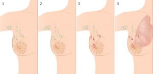
Cancer particles are carried through the bloodstream and into the lymph nodes. This entire path is divided into medicine into 4, and in some sources 6 stages (including the 0th and 6th). In the process of detecting the stage of cancer, doctors take into account:
- the size of the tumor itself;
- damage to the lymphatic system;
- the presence of metastases;
- spread of the disease to other organs.
To obtain this information, doctors use several diagnostic methods:
- Palpation. The presence of a neoplasm, its size, connection with the surrounding tissues can be detected by the doctor already upon palpation of the mammary gland.
- Ultrasound. One of the easiest ways to detect a disease. This method is relatively inexpensive, simple, painless, and quick, and therefore can be used at different ages.
- Mammography. Recommended for patients over the age of 40. It makes it possible to detect a tumor in those situations where it is difficult to feel it with your hands if there are increased calcium accumulations in the chest (this may be evidence of stage 0 cancer). The result of a mammogram is an X-ray.
- MRI. Unlike mammography, this method does not involve the use of X-ray irradiation, does not depend on the density of tissues in the mammary gland. Thus, MRI can be used at any period of the cycle and at any age. The method is called the most modern and highly informative. With its help, doctors not only learn about the size of the tumor, the peculiarities of its placement but also receive other valuable information that will be used during the operation.
- Biopsy. Most often it is used after tumor removal. Provides for a detailed study of the neoplasm under a microscope. Confirms or refutes the malignant nature of the neoplasm.
What are the early and late stages?
For the convenience of determining the treatment regimen, doctors often use the classification of breast cancer stages into:
- early;
- late.
The first is: 1, 2A, 2B, 3A. Late, respectively, are determined: 3B, 3C, 4 stages. The established stage directly affects the likelihood of complete recovery, the patient’s life expectancy, the duration of treatment, the methods of therapy used, medications, etc.

Stage 1-2 breast cancer (i.e. early stage) is treated quite successfully. The detected tumor, in this case, is removed without resection of the breast itself, that is, the woman’s breast shape is completely preserved. Chemotherapy and radiation therapy are not always used in this case. The chance of relapse after early treatment is very low.
Treatment in the later stages is complex. The breast is most often removed completely. The likelihood of a relapse is very high. A woman should undergo systematic examinations by an oncologist.
Stage 2-3 breast cancer has a ten-year patient survival rate of about 60% and 40%, respectively. At stage 4 of the disease, this figure is only 10%. Thus, the sooner a woman seeks a doctor and starts treating a disease, the more chances she has to overcome the disease.
In this case, an annual medical examination using ultrasound and mammography is mandatory for those who care about their health.
First and second stages of cancer
Stage 1 breast cancer is characterized by minor changes in the female body. The main signs of this stage are:
- the small size of the neoplasm (from a few millimeters to a maximum of 2 cm);
- absence of metastases;
- there is no prevalence of nearby tissues.
Mammography at this stage will show the presence of a neoplasm, but it is difficult to detect its malignant nature. Therefore, a fine-needle biopsy is additionally performed. A particle of the tumor is taken for further careful examination under a microscope.

If cancer is diagnosed, then the tumor must be carefully removed. In this case, as a rule, no more than 1 cm of healthy tissue is removed. After completing an additional course of chemotherapy (not always used), the patient can completely forget about the disease. Meanwhile, regular check-ups with a mammologist are mandatory.
Stage 2 breast cancer is also an early one, but the disease already requires more complex treatment. There are two types (substages) of grade 2 cancer:
- 2A. The tumor itself is still not very large, but it has a size of more than 2 cm. Cancer cells can already penetrate into the nearest lymph nodes. According to the American Cancer Society, if the lymphatic system is not affected in stage 2A, the chance of complete recovery is 85%.
- 2B. The neoplasm is up to 5 cm in size, and the disease already affects several lymph nodes.
- It is possible to identify the disease at this stage using various diagnostic methods. Often a woman herself accidentally discovers a small lump in her breast. Information on the presence of stage 2 cancer can be obtained based on blood tests for tumor markers, CT, MRI, ultrasound, and other methods.
The most effective methods of treatment at this stage can be read here.
If the lymph nodes are not affected, then at stage 2 a mastectomy (complete removal of the breast) or sectoral resection is performed (the organ is preserved, but the tumor and surrounding healthy tissues are removed). In the case when the disease has already struck the lymph nodes, they also have to be removed. In this case, oncologists call chemotherapy and radiation therapy the most effective methods of treatment.
How to distinguish and treat the third stage?
Third-degree breast cancer is also often referred to as locally advanced cancer. Most patients discover their disease at this stage. If at 1-2 stages the disease develops slowly, then already by 3 it becomes more aggressive. The main differences in the third stage are:
- the presence of a large tumor (as a rule, its diameter is more than 5 cm);
- the formation spreads deep into the tissues, the doctor can already confirm on palpation that the neoplasm extends beyond the mammary gland;
- the swelling damages the skin of the breast: it becomes flabby, may resemble a lemon peel, or is covered with sores (stage 3B);
- metastasis occurs to the nearest lymph nodes (stage 3A). The occurrence of micrometastases in other organs is possible (although at this stage it is virtually impossible to identify them); if the neoplasm enters the chest, stage 3C is diagnosed.
In the third stage of breast cancer, complex therapy should be carried out. If it is not performed, the probability of a relapse of the disease within the next 10 years is 100%. Only the removal of the tumor, in this case, is not enough.

Complete removal of the mammary gland, together with all micrometastases, shows greater effectiveness of treatment. Sometimes it is necessary to remove entire groups of lymph nodes.
The treatment regimen for the third stage is determined by the surgeon individually in each case. Most often it is supposed to use:
- preoperative therapy;
- surgical treatment;
- chemotherapy;
- hormone therapy;
- adjuvant treatment (radiation therapy).
To make the treatment more effective, the patient must systematically take blood tests for the presence of tumor markers. If the indicators do not change or increase, drugs are replaced by others.
Clinical manifestations of the fourth degree
4 degree of pathology refers to late. It is different:
- large tumor neoplasms (as a rule, it already occupies the whole mammary gland, grows into muscle tissue);
- cancer cells spread to the bones of the skeleton, metastases enter the internal organs (first to the liver and lungs, and then to the adrenal glands, brain, skin);
- close and distant lymph nodes are affected;
- the tumor itself begins to disintegrate.
As for the external manifestations of advanced cancer, we can name:
- frequent headaches;
- aching bones;
- the yellowness of all skin;
- purulent, bloody discharge from the mammary gland;
- an increase in the size of the breast itself;
- sometimes redness, swelling, and peeling of the breast;
- severe chest pain.
Although one should not hope for complete recovery at stage 4, however, oncologists say that treatment must be carried out. The main task of medicine is to relieve pain, prolong the patient’s life.
Surgical intervention is ineffective in the advanced form of the disease, but it can be performed to reduce symptoms (most often during the period of tumor decay). If metastases have gone to the lungs and liver, and the tumor itself is small, it is advisable to carry out a combined surgical intervention.

Treatment is carried out using complex therapy. The patient must use strong drugs of chemotherapy, hormone therapy (if appropriate). In the case when a woman is sensitive to targeted therapy, she is also performed.
Radiation therapy is also an integral part of the treatment. It is especially effective in cases where metastases are rare.
Long-term treatment, even at this stage, gives about 10% of women a chance to continue life for more than 5 years.